

Kidney stones are one of the most common urological conditions seen across all age groups. Many small stones pass on their own with hydration, time, and medication. However, when a stone is causing persistent pain or blockage, expert intervention becomes necessary. Extracorporeal Shock Wave Lithotripsy (ESWL) represents the most established non-invasive therapeutic option available for eligible patients.
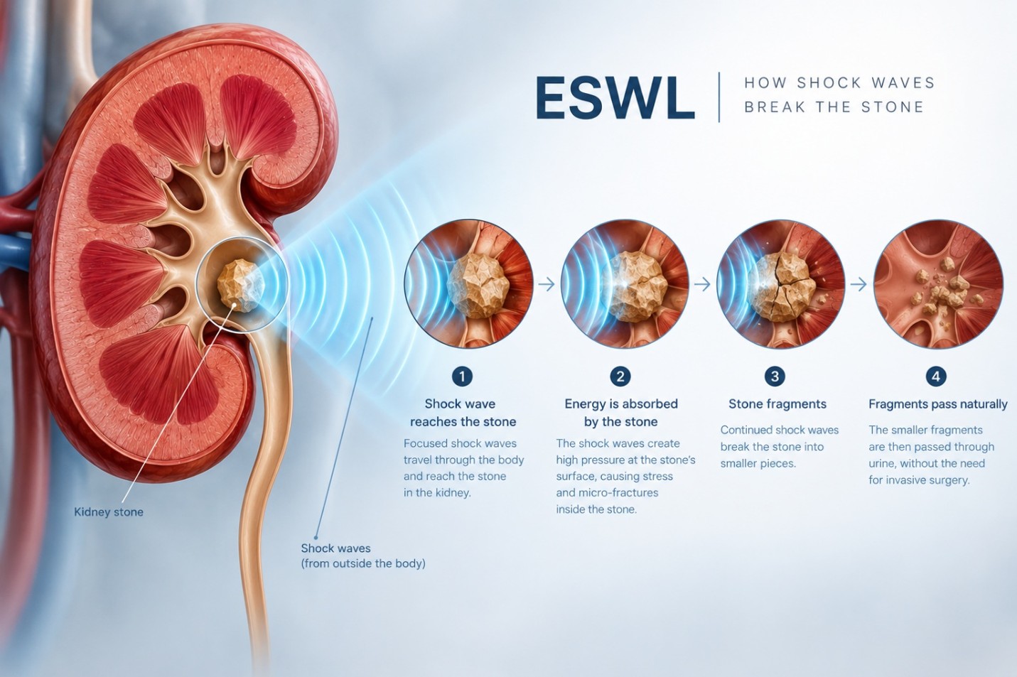
ESWL utilizes focused shock waves generated outside the body to break kidney stones into smaller fragments. Once fragmented, these pieces are sufficiently reduced in size to pass through the urinary tract naturally, typically over the course of several days.
In accordance with clinical guidelines from the European Association of Urology (EAU) and the American Urological Association (AUA), ESWL is recommended within the following parameters:
Precise localization of the calculus is fundamental to the efficacy of ESWL. Targeting is routinely conducted via X-ray, ultrasound (US), or a combined dual-imaging approach:
Because each modality exhibits inherent limitations, employing a dual-imaging approach (simultaneous X-ray and ultrasound) optimizes stone tracking throughout the treatment cycle. This combined visualization supports higher stone-free success rates while minimizing procedural complications.
Suitability for ESWL is evaluated based on several interdependent factors:
ESWL is not universally indicated. The procedure may not be recommended in clinical scenarios involving pregnancy, uncorrected bleeding diatheses, uncontrolled urinary tract infections, or specific stone compositions known to resist shock wave fragmentation. Final clinical selection must always be determined by a qualified physician following a comprehensive diagnostic workup.
The objective of ESWL is the disintegration of a calculus into fragments small enough to traverse the urinary tract spontaneously. Fragmentation efficacy directly correlates with the total cumulative shock wave energy dose administered during a treatment session.
Delivering a well-judged, sufficient energy dose is critical during treatment planning. Adequate fragmentation and clearance significantly mitigate the requirement for secondary interventions, such as repeat ESWL, ureteroscopy (URSL), or percutaneous nephrolithotomy (PCNL).
Following ESWL, patients commonly experience mild discomfort as stone gravel passes through the urinary tract. Clinicians routinely prescribe appropriate analgesia and advise increased oral fluid intake to facilitate clearance.
The primary reason patients and clinicians select ESWL is its completely non-invasive nature, eliminating surgical incisions and general operative trauma. Furthermore, this non-invasive profile makes ESWL one of the preferred modalities for eligible paediatric populations. Overall recovery duration, procedural tolerance, and the total number of sessions remain case-dependent, requiring individualized consultation between the treating physician and the patient.
Healthware serves as the solution partner for Dornier-manufactured ESWL systems for hospitals and urology centres. Both platforms incorporate electromagnetic shock wave generators, engineered for stable, reproducible shock wave administration.
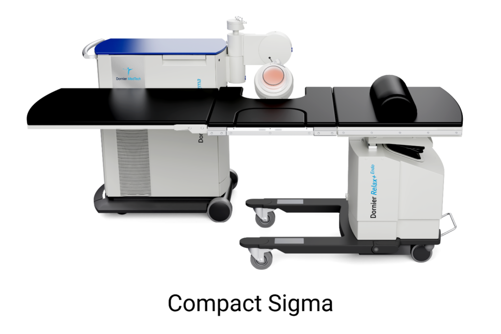
1. Dornier Compact Sigma
A modular lithotripter engineered with a 150 mm penetration depth, pairing a multi-functional patient table with an X-ray C-arm. Its isocentric architecture ensures the shock wave source and imaging apparatus remain continuously aligned around a single focal point, supporting simultaneous X-ray and ultrasound localization.
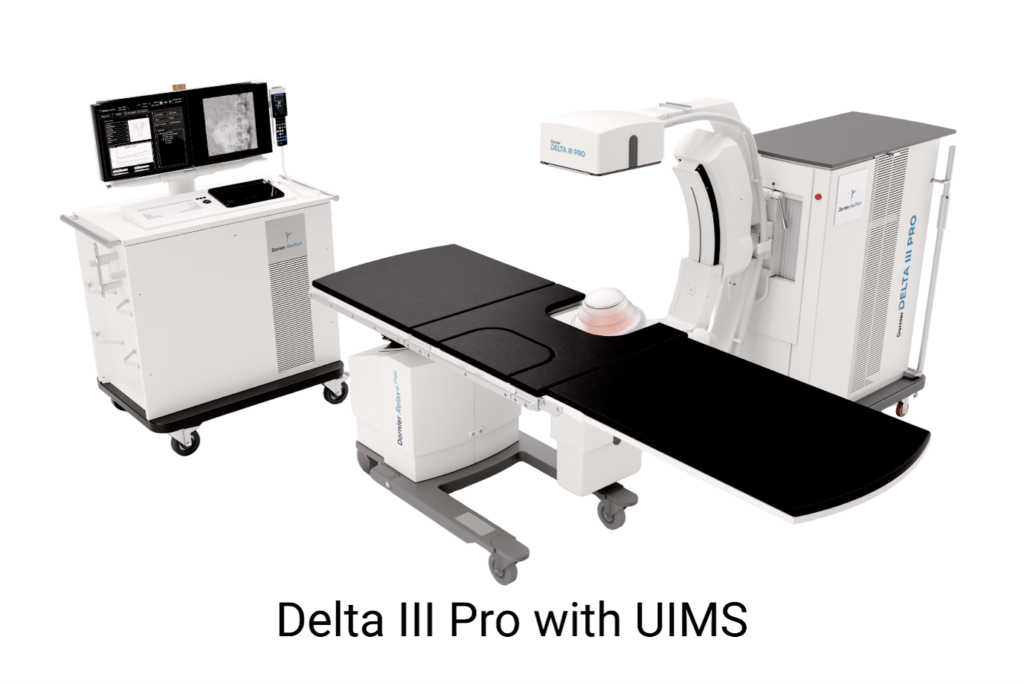
2. Dornier Delta III Pro
An integrated lithotripter providing dual imaging modalities (ultrasound and fluoroscopy) with an extended 170 mm focal depth, an isocentric ultrasound arm, and specialized interface monitoring software.
Building upon the Compact Sigma feature set, the Delta III Pro incorporates:
Kidney stone management must always be directed by a qualified urologist or nephrologist. Specialized clinicians evaluate stone size, location, and composition within the context of the patient’s comprehensive medical history. Diagnostic modalities—including ultrasound, CT imaging, and comprehensive blood and urine analyses—are essential to determine whether conservative management, ESWL, or surgical intervention constitutes the appropriate clinical pathway.
ESWL continues to expand therapeutic possibilities for patients with urolithiasis by providing an effective non-surgical pathway for qualified candidates. It is not an all-inclusive solution, nor does it replace thorough clinical and diagnostic evaluation. Patients presenting with symptoms indicative of kidney stones should seek immediate consultation with a qualified urologist.
This article is intended for general informational purposes only and does not constitute formal medical advice, diagnosis, or treatment. It must not be utilized as a substitute for professional clinical evaluation. Individuals experiencing symptoms should consult a licensed healthcare professional.
Content last reviewed: 29-07-2026.